Application
An inflation system to mimic the air variation of the human lungs and asses respiration-induced MRI artefacts.
Contributors
Philippe De Tillieux1, Ryan Topfer1, Alexandru Foias1, Iris Leroux1, Imanne El Maachi1, Hugues Leblond1, Nikola Stikov1,3 and Julien Cohen-Adad1,4
Estimated cost
$400 CAD
Progress
Hardware: Prototype, MIT License.
Software: Alpha version, MIT License.
The aim of this project is to provide a compatible MRI phantom that will simulate the respiration of a human subject in an automated way. A pressure probe is used in order to communicate in continuous time to Matlab the evolution of the respiratory cycle of the subject.
The complete design has been divided into three main components: the inflation system, the phantom, and the respiratory sensor:
- the inflation system consists of two solenoid valves which connect the phantom to a compressed air tank and a vacuum pump.
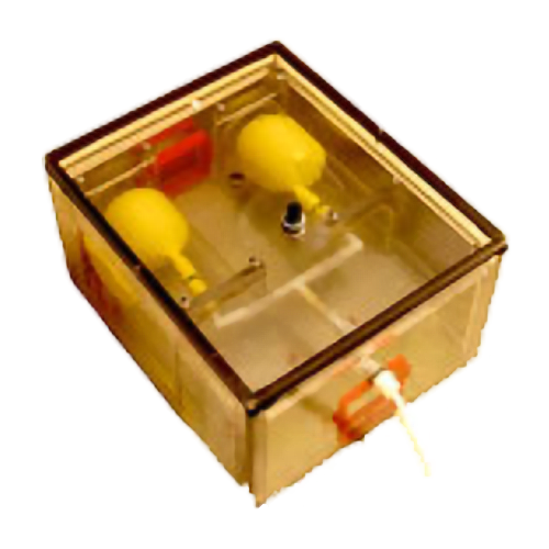
- the phantom comprises a box containing two mechanically resistant “punch‐balloon” lungs, and an ex-vivo human spine preserved by conservation liquid.
- the respiratory sensor allows real‐time tracking of the pressure inside the tubes and to control both the positive and negative pressure valve opening. It is connected to the phantom and to the tube linking the inflation system by a T‐type connector.
The phantom mimics the change in air volume and can thus serve as a testing platform for the measurement and correction of respiratory motion artifacts during spinal cord imaging. Its simplicity and size allow it to be easily adapted to other applications.
Publications
Affiliations
1NeuroPoly Lab, Institute of Biomedical Engineering, Polytechnique Montreal, Montreal, Quebec, Canada;
2Department of Anatomy, Université du Québec à Trois‐RivièresTrois‐Rivières, Quebec, Canada;
3Montreal Heart Institute, Université de Montréal, Montreal, Quebec,Canada;
4Functional Neuroimaging Unit, CRIUGM, Université de Montréal, Montreal, Quebec, Canada.

